HBOT for Radiation Necrosis and Late Radiation Tissue Injury

Key Takeaways
- Late radiation tissue injury can appear months or years after radiotherapy. It is driven by progressive damage to small blood vessels, chronic inflammation, fibrosis, reduced tissue oxygenation and impaired healing. It can affect the bladder, rectum and bowel, jaw and other bones, skin and soft tissues, breast and chest wall, head and neck structures, and the brain.1 2 3
- The term radiation necrosis covers several clinically different problems. Soft-tissue radionecrosis, osteoradionecrosis of bone and cerebral radiation necrosis should not be treated as though they have the same evidence base or the same care pathway.
- Hyperbaric oxygen therapy (HBOT) is a physician-prescribed treatment in which a patient breathes near-100% oxygen in a medical hyperbaric chamber. A common course for late radiation injury is 30-40 sessions, usually five days per week. Oxygen exposure is often around 80-90 minutes, while the full chamber visit is commonly 90-120 minutes. Protocols must be individualised.1 4 5
- The strongest randomised evidence supports HBOT for radiation-induced cystitis. Evidence for chronic radiation proctitis is mixed but supports consideration in selected, refractory cases.1 3 6
- Emerging evidence also suggests a possible role in vaginal late radiation tissue injury after pelvic radiotherapy. In a 2025 retrospective series of 19 patients, 15 (79%) improved in at least one symptom after HBOT, but the study was small and uncontrolled, so the finding is encouraging rather than definitive.7
- For osteoradionecrosis of the jaw, HBOT may have a selective perioperative role, but modern trials and a 2024 international guideline do not support routine prophylactic or blanket use.8 9 10
- For cerebral radiation necrosis, published HBOT evidence remains limited to small, mainly retrospective series. HBOT may be discussed for carefully selected patients within a neuro-oncology pathway, but it is not an established first-line treatment and must not delay investigation for tumour recurrence.1 11 12
- HBOT is generally well tolerated in appropriately selected patients, but it is not risk-free. Ear barotrauma and temporary short-sightedness are the most recognised adverse effects; oxygen-induced seizures and pulmonary barotrauma are rare.1 3
- HBOT should be used as an adjunct within multidisciplinary cancer survivorship care, not as a replacement for oncology, surgery, urology, gastroenterology, gynaecological oncology, oral and maxillofacial, wound-care or neuro-oncology treatment.
Quick Summary
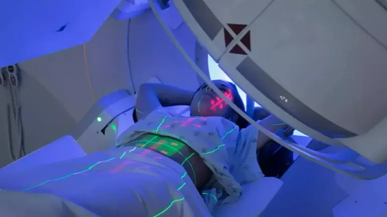
Radiotherapy is an established cancer treatment, but healthy tissue within or close to the treatment field may develop delayed injury. Unlike acute side effects, which usually occur during treatment or shortly afterwards, late radiation tissue injury can emerge after a symptom-free interval and may continue to progress.
The underlying problem is often described as a chronic wound-healing disorder. Radiation can damage vascular endothelial cells and reduce the density of small blood vessels. Over time, tissue becomes relatively hypovascular, hypoxic and fibrotic, with reduced capacity to repair after minor trauma, infection or surgery. In severe cases, ulceration, fistula formation, non-healing wounds, bleeding, exposed bone or tissue necrosis can develop.1 2 3
Symptoms depend on the site involved. They may include visible blood in the urine, urinary urgency or pain; rectal bleeding, urgency or bowel pain; vaginal pain, dyspareunia, dryness, bleeding, ulceration or stenosis; non-healing oral wounds, exposed jaw bone or jaw pain; skin ulceration, fibrosis or reduced movement; and, in cerebral radiation necrosis, headaches, seizures, cognitive change, weakness or other neurological symptoms.
These symptoms are not specific to radiation injury. Recurrent cancer, infection, vascular disease, medication effects and unrelated conditions may present in similar ways. A diagnosis should therefore be made by the relevant specialist team before HBOT is considered. New neurological symptoms, heavy bleeding, sepsis, severe pain, obstruction, rapidly progressive swelling or a non-healing wound with concern for recurrence require urgent assessment.
HBOT is used because it can markedly increase dissolved oxygen in plasma and improve the oxygen gradient into poorly perfused tissue. Repeated treatments may stimulate new microvascular growth and support oxygen-dependent repair processes. The biological rationale is strong, but clinical benefit is site-specific and the certainty of evidence varies substantially.1 2 3
What Is Radiation Necrosis?
Radiation necrosis is one end of a spectrum of late radiation tissue injury. The preferred clinical description often depends on the tissue involved:
- Soft-tissue radionecrosis affects irradiated skin, subcutaneous tissue, mucosa or internal organs. Examples include radiation cystitis, radiation proctitis, vaginal ulceration, fibrosis or stenosis after pelvic radiotherapy, laryngeal injury, chest-wall or breast fibrosis, pelvic wounds and ulceration.
- Osteoradionecrosis affects irradiated bone, most commonly the mandible after head and neck radiotherapy. It may present with exposed bone, pain, infection, fistula formation, fracture or failure of a surgical wound to heal.
- Cerebral radiation necrosis is delayed injury to brain tissue after cranial radiotherapy or stereotactic radiosurgery. It is clinically distinct because imaging changes can closely resemble tumour progression and treatment decisions often depend on advanced imaging, serial scans or tissue diagnosis.
The biology is complex. Radiation-related endothelial injury, capillary loss and impaired perfusion create chronic hypoxia. Persistent inflammatory and fibrogenic signalling promotes fibrosis and reduces regenerative capacity. Tissue may remain stable for years and then break down after dental extraction, surgery, infection, pressure, friction or another relatively small insult.1 2 3
What Is HBOT?
Hyperbaric oxygen therapy is a medical treatment in which patients breathe near-100% oxygen at a pressure greater than normal atmospheric pressure inside a purpose-built chamber. In evidence-based medical practice, treatments for late radiation injury are commonly delivered at approximately 2.0-2.5 atmospheres absolute. This is different from so-called mild hyperbaric services, which should not be assumed to provide the same dose or clinical effect.1 4 5
At pressure, much more oxygen dissolves directly in plasma. This allows oxygen to travel beyond the limits imposed by damaged microcirculation and increases the diffusion gradient into hypoxic tissue. A single exposure improves oxygen availability temporarily. The intended therapeutic effect in late radiation injury comes from a repeated course that supports vascular and tissue remodelling over time.
A typical course is around 30-40 treatments, delivered once daily on weekdays. Some patients need fewer or more sessions, and selected surgical pathways may use treatments before and after an operation. The pressure, oxygen time, use of air breaks and total number of sessions should be prescribed by a hyperbaric physician according to the anatomical site, severity, treatment objective, response and relevant comorbidities.1 3 5
Biological Rationale for HBOT
HBOT does not remove radiation from tissue and it does not reverse every structural change caused by radiotherapy. Its role is to improve the biological environment for repair. Repeated treatment may help through several linked mechanisms:
- Increasing tissue oxygen availability. Higher plasma oxygen levels improve diffusion into areas with compromised blood flow and may temporarily support cells at the edge of an ischaemic or necrotic area.
- Promoting angiogenesis and neovascularisation. Repeated oxygen exposure can stimulate signalling involved in the formation and maturation of new microvessels, helping to improve perfusion of chronically hypoxic tissue.
- Supporting fibroblasts, collagen deposition and epithelial repair. These oxygen-dependent processes are important for wound closure, mucosal recovery and healing around surgery.
- Modulating oedema and inflammation. Hyperoxia causes vasoconstriction in normally perfused vessels while maintaining oxygen delivery, which may reduce oedema. It can also influence inflammatory signalling and leukocyte-endothelial interactions.
- Supporting repair in irradiated bone and soft tissue. Where surgery is required, improved oxygenation may help the surrounding tissue tolerate debridement, reconstruction or wound closure.
These mechanisms are biologically plausible across many tissues, but biological plausibility is not the same as proof of clinical benefit. Treatment decisions should therefore be based on the evidence for the particular organ and clinical problem, rather than on mechanism alone.1 2 3
What Does the Evidence Show?
1. Radiation-Induced Cystitis: Strongest Evidence
Radiation cystitis is the clearest evidence-supported use of HBOT within late radiation tissue injury. Patients may experience recurrent haematuria, urgency, frequency, pain and reduced bladder capacity. Before HBOT, bleeding should be assessed by urology and other causes, including recurrent malignancy, infection, stones and medication-related bleeding, should be addressed.
The landmark RICH-ART randomised trial included 87 patients with chronic radiation cystitis. HBOT was delivered over 30-40 sessions at 2.4-2.5 atmospheres absolute. At 6-8 months, improvement in patient-reported urinary symptoms was greater with HBOT than with standard care, with a between-group difference of 10.1 points on the EPIC urinary score and a reported number needed to treat of approximately three.1 13
Five-year follow-up showed that improvement in urinary and bowel symptom scores was maintained in the treated cohort. Because nearly all control participants later crossed over to HBOT, the long-term study demonstrates durability after treatment rather than a five-year randomised comparison. Some patients with recurrent symptoms received an additional course.1 14
Taken together with systematic reviews, meta-analyses and real-world cohorts, the evidence supports HBOT as a credible bladder-preserving adjunct for appropriately selected patients with persistent or recurrent radiation cystitis. It should still sit within a urology-led pathway, particularly where there is severe bleeding, clot retention, anaemia or concern about recurrence.
2. Radiation Proctitis and Chronic Pelvic Bowel Injury: Mixed Evidence
Chronic radiation proctitis or proctopathy may cause rectal bleeding, urgency, tenesmus, pain, incontinence and ulceration. The diagnosis usually requires specialist gastrointestinal assessment because symptoms may have multiple causes and endoscopic procedures in heavily irradiated tissue require appropriate expertise.
Two sham-controlled trials reached different conclusions. In HORTIS-IV, 120 evaluable patients with refractory radiation proctitis had greater improvement after HBOT than after sham treatment, with an absolute risk reduction of 32% and a number needed to treat of approximately three. Improvements in bowel-specific quality of life were also reported.15
The later HOT2 phase 3 trial included 84 patients with chronic bowel dysfunction after pelvic radiotherapy and found no clinically relevant benefit at 12 months. Differences in patient selection, symptom severity, time from radiotherapy and outcome measurement may partly explain the conflicting results.6
The balanced conclusion is that HBOT may help selected patients with refractory, objectively supported radiation proctitis, but benefit should not be promised and referral criteria matter. Gastroenterology or colorectal review, optimisation of conventional treatment and careful selection are essential.
3. Osteoradionecrosis of the Jaw: A Selective, Multidisciplinary Role
Osteoradionecrosis of the jaw is a serious complication of head and neck radiotherapy. Management may include specialist dental care, treatment of infection, analgesia, conservative wound care, debridement, resection and reconstruction. The role of HBOT has changed as radiotherapy, dental prevention, antibiotics and reconstructive surgery have evolved.
For prevention around dental extraction or implant surgery, the HOPON randomised trial found osteoradionecrosis in 6% of both the HBOT and control groups at six months. The trial stopped early because the overall event rate was lower than expected, but its findings do not support routine HBOT for every dentoalveolar procedure in an irradiated mandible.8
For established mandibular osteoradionecrosis requiring surgery, combined data from the DAHANCA-21 and NWHHT2009-1 trials showed healing in 70% of patients who received surgery plus HBOT and 51% who received surgery alone. The difference was not statistically significant and the trials were underpowered, so the result is suggestive rather than definitive.9
The 2024 ISOO-MASCC-ASCO guideline concluded that evidence for HBOT in prevention and management of jaw osteoradionecrosis remains limited and largely does not justify routine use. The Cochrane review found a small improvement in pain but low or uncertain evidence for several other outcomes.3 10
HBOT may still be considered for selected patients, especially as an adjunct to surgery where local tissue quality is poor, healing has failed or a specialist oral and maxillofacial team believes the potential benefit outweighs the burden and risk. It should not be presented as a mandatory universal protocol or as a stand-alone substitute for definitive surgical care.
4. Breast, Chest Wall, Head and Neck, and Other Soft-Tissue Injury
HBOT has been used for painful fibrosis, oedema, restricted movement, ulceration and non-healing wounds after breast, chest-wall, head and neck, gynaecological and other radiotherapy. Most published evidence is observational, and the clinical problems grouped under soft-tissue radionecrosis are highly varied.
The 2024 HONEY randomised clinical trial studied women with late local breast or chest-wall toxicity. In the intention-to-treat analysis, HBOT did not significantly reduce pain, although fibrosis improved. Among women who completed HBOT, both pain and fibrosis improved more than in controls, but this subgroup analysis is vulnerable to selection effects because many participants allocated to HBOT declined the demanding treatment schedule.1 16
For laryngeal, cutaneous, pelvic and other soft-tissue necrosis, case series and mixed-site cohorts report healing or symptom improvement in some patients, particularly where tissue is ulcerated or surgery is planned. High-quality site-specific trials are scarce.1 2 3
HBOT may be reasonable when there is a clearly defined radiation-related wound-healing problem, conventional measures have been optimised and the treatment objective is measurable, such as wound closure, reduced bleeding, improved function or safer reconstruction.
5. Vaginal Late Radiation Tissue Injury: Encouraging but Preliminary Evidence
Late radiation tissue injury of the vagina may follow pelvic radiotherapy for cervical, anal, rectal or other gynaecological malignancies. Symptoms can include dyspareunia, pain, dryness, bleeding, discharge, ulceration or necrosis, fibrosis, shortening, adhesions and stenosis. These problems may substantially affect quality of life and sexual function. Assessment should involve gynaecological oncology or a specialist pelvic late-effects service, with recurrent malignancy, infection and fistula excluded before symptoms are attributed to radiation injury.7
In 2025, Möring and colleagues published a retrospective analysis of 19 patients referred for HBOT because vaginal symptoms were their primary late radiation complaint. Patients with vesicovaginal or rectovaginal fistulas, predominantly non-vaginal symptoms or fewer than 20 completed sessions were excluded. The median age was 42 years, cervical cancer was the most common primary diagnosis, and patients received a median of 40 sessions (range 30-50), using 80 minutes of 100% oxygen at 2.5 atmospheres absolute.7
Fifteen of 19 patients (79%) met the study's responder definition, which required improvement or resolution of at least one vaginal symptom, and the median number of symptoms improved was three. Among patients with paired before-and-after data, dyspareunia improved in 9 of 11 (82%), pain in 10 of 14 (71%), self-reported anatomical changes in 8 of 10 (80%) and bleeding in 7 of 11 (64%). On examination, ulceration or necrosis improved in 8 of 9 patients (89%), with complete resolution in 4 of 9. Most responders with three-month data maintained the benefit; one patient worsened in association with infection.7
No major adverse events were reported. Mild fatigue affected 53%, transient myopia 32%, and three patients experienced difficulty equalising ear pressure. Some quality-of-life and sexual-function measures improved, but data were incomplete and not all outcomes moved in the same direction.7
The study is encouraging but cannot establish that HBOT caused the improvements. Many participants had already used dilators, pelvic-floor physiotherapy or hormone treatment, reinforcing that HBOT should be considered as an adjunct within a broader gynaecological late-effects plan rather than a replacement for established care.7
For a patient with persistent, objectively assessed vaginal ulceration, bleeding, pain, dyspareunia or fibrosis after conventional measures have been reviewed, HBOT may be reasonable to discuss. The expected benefit, uncertainty, treatment burden and measurable goals should be agreed in advance.
6. Cerebral Radiation Necrosis: Promising but Unproven
Cerebral radiation necrosis can occur after conventional cranial radiotherapy or stereotactic radiosurgery. Symptoms may result from local tissue injury and surrounding oedema and can include headache, seizures, weakness, speech or visual change, cognitive decline and altered behaviour.
The central clinical challenge is diagnosis. Radiation necrosis and tumour progression can look similar on standard MRI and may coexist. Assessment may require serial MRI, perfusion imaging, spectroscopy, amino-acid PET or surgery and histology, depending on the case. HBOT should never be started in a way that delays this evaluation.12
For symptomatic cerebral radiation necrosis, corticosteroids are commonly used first to reduce oedema. In steroid-refractory disease, bevacizumab has the strongest published evidence, while laser interstitial thermal therapy, surgical resection and other approaches may be appropriate according to lesion location, severity, diagnostic uncertainty and the patient's wider oncology plan. International stereotactic radiosurgery recommendations list HBOT as a possible option in selected cases, not as the default treatment.12
Published HBOT studies for brain radiation necrosis are small and mainly retrospective or uncontrolled. Reviews describe clinical or radiological improvement in some patients, but the evidence is insufficient for a clear general recommendation.1 11 17
A carefully selected patient with persistent symptoms, limited alternatives or contraindications to other treatments may reasonably be discussed by neuro-oncology, radiation oncology, neurosurgery and hyperbaric medicine. The uncertainty, time commitment and alternative treatments should be explained clearly.
HBOT Care Strategy
HBOT is most useful when it is integrated into a defined multidisciplinary plan. A safe and clinically meaningful pathway should include the following steps:
- Confirm the diagnosis and anatomical site. Previous radiotherapy alone is not enough to establish radiation necrosis. The team should identify objective evidence of tissue injury and exclude recurrent cancer, infection and other treatable causes.
- Stabilise urgent complications first. Major haemorrhage, clot retention, sepsis, airway compromise, bowel obstruction, pathological fracture, rapidly progressive neurological deficit or uncontrolled pain requires urgent conventional assessment and treatment.
- Review standard treatment already given. HBOT may be considered when symptoms are persistent, recurrent or refractory, or when it is being used to support a planned operation in compromised tissue. It should not replace a necessary procedure.
- Define a measurable goal. Depending on the site, this might be reduced haematuria or transfusion need, improved urinary or bowel symptom scores, reduced vaginal bleeding or pain, healing of a vaginal ulcer, improved dyspareunia, wound closure, reduced exposed bone, improved mouth opening, reduced fibrosis, improved mobility or a radiological and steroid-sparing response in brain necrosis.
- Assess fitness for HBOT. The hyperbaric physician should review ear and sinus function, lung disease and thoracic imaging where indicated, seizure history, diabetes, implanted devices, pregnancy status, current chemotherapy drugs and other medicines, mobility and ability to attend a multi-week course.
- Individualise the protocol. A common late-radiation course is 30-40 weekday sessions at around 2.0-2.5 atmospheres absolute, but dose and duration vary. Some surgical protocols add postoperative sessions. Response should be reviewed during and after treatment rather than assuming that every patient needs the same number.
- Coordinate follow-up. Improvement may continue after the course has finished. Persistent or recurrent symptoms should prompt reassessment, not an automatic assumption that more HBOT is required.
Safety and Patient Experience
HBOT is generally well tolerated when delivered in an appropriately regulated medical facility with trained staff, but it has practical and medical burdens. A course usually requires weekday attendance for six to eight weeks, and each visit may occupy around two hours once compression, oxygen periods, air breaks and decompression are included.
The most common problems are difficulty equalising middle-ear pressure, ear discomfort and minor middle-ear barotrauma. Temporary short-sightedness can develop during a long course and usually resolves after treatment. Sinus discomfort, fatigue and claustrophobia can also occur.1 3
Oxygen-induced seizures are rare, reported at roughly one event per 2,000-3,000 treatments in the 2026 clinical review. Pulmonary barotrauma and pneumothorax are also rare but are relevant in patients with certain lung conditions.1
Medication interactions, recent surgery, implanted devices and current or previous anticancer drugs should be reviewed by the treating physician. Patients should not stop oncology medication without advice from their oncology team.
Available reviews have not shown that HBOT promotes tumour growth or recurrence. Even so, suspected active or recurrent cancer must be investigated because the symptoms attributed to radiation injury may instead reflect malignancy, and the overall treatment plan must remain oncology-led.18
Summary
Hyperbaric oxygen therapy has a credible role in the management of selected late radiation injuries, but the phrase radiation necrosis should not be treated as a single diagnosis with a single level of evidence.
The strongest evidence supports HBOT for radiation-induced cystitis. Chronic radiation proctitis has conflicting randomised evidence but remains a reasonable consideration in selected refractory cases. For vaginal late radiation tissue injury, a 2025 retrospective series reported improvement in 79%, but the sample was small and uncontrolled, so the evidence remains preliminary. For jaw osteoradionecrosis, routine prophylactic use is not supported by modern evidence, while a selective perioperative role may remain appropriate in specialist practice. For breast, chest wall and other soft-tissue injuries, evidence is suggestive but heterogeneous. For cerebral radiation necrosis, HBOT remains an option with limited evidence and should be considered only within a neuro-oncology pathway.
For the right patient, HBOT may improve symptoms, quality of life and tissue healing, or support surgery in an irradiated field. It cannot guarantee recovery, replace necessary conventional treatment or remove the need to exclude recurrent cancer.
Every patient should receive an individual assessment by a physician trained in hyperbaric medicine, with input from the relevant oncology and specialists. The decision should balance the strength of evidence for that anatomical site, clinical severity, alternative treatments, treatment burden, medical risk and the patient's goals.
Medical Disclaimer
This article is for general information and does not replace individual medical advice, diagnosis or emergency care. Treatment decisions should be made by the patient's clinical team after review of the diagnosis, cancer status, comorbidities, medicines and available alternatives.
References
Footnotes
- Dejonckheere CS, Käsmann L, Schmeel LC, et al. Hyperbaric oxygen therapy for chronic radiotherapy-related adverse effects: a clinically focused review. CA Cancer J Clin. 2026;76(1):e70058. doi:10.3322/caac.70058. ↩ ↩2 ↩3 ↩4 ↩5 ↩6 ↩7 ↩8 ↩9 ↩10 ↩11 ↩12 ↩13 ↩14 ↩15 ↩16 ↩17 ↩18
- Cuccia F, Neville Cracchiolo A, Piras A, et al. Between hope and uncertainty: the elusive evidence on hyperbaric oxygen therapy and radiotherapy. Support Care Cancer. 2026;34(2):157. doi:10.1007/s00520-026-10323-8. ↩ ↩2 ↩3 ↩4 ↩5 ↩6
- Lin ZC, Bennett MH, Hawkins GC, et al. Hyperbaric oxygen therapy for late radiation tissue injury. Cochrane Database Syst Rev. 2023;8(8):CD005005. doi:10.1002/14651858.CD005005.pub5. ↩ ↩2 ↩3 ↩4 ↩5 ↩6 ↩7 ↩8 ↩9 ↩10 ↩11
- Mathieu D, Marroni A, Kot J. Tenth European Consensus Conference on Hyperbaric Medicine: recommendations for accepted and non-accepted clinical indications and practice of hyperbaric oxygen treatment. Diving Hyperb Med. 2017;47(1):24-32. doi:10.28920/dhm47.1.24-32. ↩ ↩2
- Undersea and Hyperbaric Medical Society. Indications for hyperbaric oxygen therapy: delayed radiation injury (soft tissue and bony necrosis). UHMS; 2020 cited 2026 Aug. ↩ ↩2 ↩3
- Glover M, Smerdon GR, Andreyev HJ, et al. Hyperbaric oxygen for patients with chronic bowel dysfunction after pelvic radiotherapy (HOT2): a randomised, double-blind, sham-controlled phase 3 trial. Lancet Oncol. 2016;17(2):224-233. doi:10.1016/S1470-2045(15)00461-1. ↩ ↩2
- Möring MM, Valkenburg AC, Schuur-van't Hof N, van Beekhuizen HJ, Lansdorp CA. Reduced symptoms of late radiation tissue injury of the vagina after treatment with hyperbaric oxygen therapy: a retrospective analysis of 19 patients. Gynecol Oncol. 2025;197:27-33. doi:10.1016/j.ygyno.2025.04.003. ↩ ↩2 ↩3 ↩4 ↩5 ↩6
- Shaw RJ, Butterworth CJ, Silcocks P, et al. HOPON: a randomized controlled trial of hyperbaric oxygen to prevent osteoradionecrosis of the irradiated mandible after dentoalveolar surgery. Int J Radiat Oncol Biol Phys. 2019;104(3):530-539. doi:10.1016/j.ijrobp.2019.02.044. ↩ ↩2
- Forner LE, Dieleman FJ, Shaw RJ, et al. Hyperbaric oxygen treatment of mandibular osteoradionecrosis: combined data from the DAHANCA-21 and NWHHT2009-1 randomized clinical trials. Radiother Oncol. 2022;166:137-144. doi:10.1016/j.radonc.2021.11.021. ↩ ↩2
- Peterson DE, Koyfman SA, Yarom N, et al. Prevention and management of osteoradionecrosis in patients with head and neck cancer treated with radiation therapy: ISOO-MASCC-ASCO guideline. J Clin Oncol. 2024;42(16):1975-1996. doi:10.1200/JCO.23.02750. ↩ ↩2
- Co J, De Moraes MV, Katznelson R, et al. Hyperbaric oxygen for radiation necrosis of the brain. Can J Neurol Sci. 2020;47(1):92-99. doi:10.1017/cjn.2019.290. ↩ ↩2
- Vellayappan B, Lim-Fat MJ, Kotecha R, et al. A systematic review informing the management of symptomatic brain radiation necrosis after stereotactic radiosurgery and International Stereotactic Radiosurgery Society recommendations. Int J Radiat Oncol Biol Phys. 2024;118(1):14-28. doi:10.1016/j.ijrobp.2023.07.015. ↩ ↩2 ↩3
- Oscarsson N, Müller B, Rosén A, et al. Radiation-induced cystitis treated with hyperbaric oxygen therapy (RICH-ART): a randomised, controlled, phase 2-3 trial. Lancet Oncol. 2019;20(11):1602-1614. doi:10.1016/S1470-2045(19)30494-2. ↩
- Oscarsson N, Rosén A, Müller B, et al. Radiation-induced cystitis treated with hyperbaric oxygen therapy (RICH-ART): long-term follow-up of a randomised, controlled, phase 2-3 trial. EClinicalMedicine. 2025;83:103214. doi:10.1016/j.eclinm.2025.103214. ↩
- Clarke RE, Tenorio LMC, Hussey JR, et al. Hyperbaric oxygen treatment of chronic refractory radiation proctitis: a randomized and controlled double-blind crossover trial with long-term follow-up. Int J Radiat Oncol Biol Phys. 2008;72(1):134-143. doi:10.1016/j.ijrobp.2007.12.048. ↩
- Mink van der Molen DR, Batenburg MCT, Maarse W, et al. Hyperbaric oxygen therapy and late local toxic effects in patients with irradiated breast cancer: a randomized clinical trial. JAMA Oncol. 2024;10(4):464-474. doi:10.1001/jamaoncol.2023.6776. ↩
- Hajikarimloo B, Kavousi S, Jahromi GG, et al. Hyperbaric oxygen therapy as an alternative therapeutic option for radiation-induced necrosis following radiotherapy for intracranial pathologies. World Neurosurg. 2024;186:51-61. doi:10.1016/j.wneu.2024.01.161. ↩
- Feldmeier J, Carl U, Hartmann K, Sminia P. Hyperbaric oxygen: does it promote growth or recurrence of malignancy? Undersea Hyperb Med. 2003;30(1):1-18. PMID:12841604. ↩